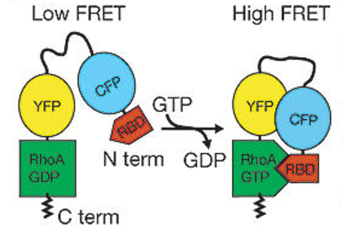
In this biosensor, an affinity reagent (RBD, an effector fragment) binds selectively to the active conformatoin of RhoA. RBD binding to RhoA changes the orientation and proximity of the YFP and CFP, producing a FRET signature for the activated state. Note that interactions with GDI remain intact because the C-terminus has not been altered.
References
Zawistowski, J.S., Sabouri-Ghomi, M., *Danuser, G., *Hahn, K.M. and *Hodgson, L. A RhoC biosensor reveals differences in the activation kinetics of RhoA and RhoC in migrating cells. PLoS One, 8(11): e79877, 2013. PMC3818223. Online article | Free PMC article
Pertz, O., Hodgson, L., Klemke, R.R., and Hahn, K.M. Spatio-temporal dynamics of RhoA activity in migrating cells. Nature, 440(7087):1069-1072, 2006. Online article | PDF
Methods articles
Hodgson, L., Pertz, O., and Hahn, K.M. Design and Optimization of genetically encoded fluorescent biosensors: GTPase biosensors. Methods Cell Biol, 85:63-81, 2008. [Link downloads PDF] Online article
*Note: this older methods article contains a useful description of biosensor image analysis.
Hodgson, L. Nalbant, P. Shen, F., and Hahn, K. Imaging and photobleach correction of Mero-CBD, sensor of endogenous Cdc42 activation. Methods Enzymol., 406:140-156, 2006. Online article | PDF
*Note: this article is focused on dye-based biosensors, but includes a method to correct for bleaching of any fluorophore.
~ Updated 04/12/2021